دوتنه:PET-MIPS-anim.gif
د همدې ليدنې کچه: ۳۹۸ × ۶۰۰ پېکسل. نورې ژورليدنې: ۱۵۹ × ۲۴۰ پېکسل | ۴۴۶ × ۶۷۲ پېکسل.
اصلي دوتنه (۴۴۶ × ۶۷۲ پېکسل, د دوتنې کچه: ۱٫۶۵ مېگابايټ, MIME بڼه: image/gif، حلقه، ۳۲ چوکاټونه، ۶٫۴ s)
لنډيز
| څرگندونه |
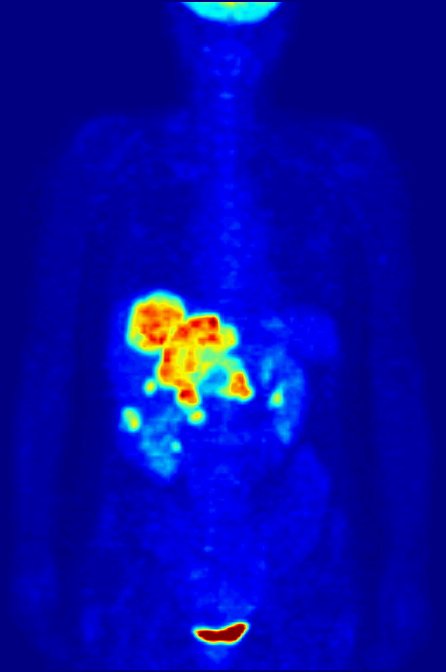
English: Maximum Intensity Projection (MIP) of a wholebody positron emission tomography (PET) acquisition of a 79 kg (174 lb) weighting female after intravenous injection of 371 MBq of 18F-FDG (one hour prior measurement). The investigation has been performed as part of a tumor diagnosis prior to applying a radiotherapy (tumor staging step). Besides normal accumulation of the tracer in the heart, bladder, kidneys and brain, liver metastases of a colorectal tumor are clearly visible within the abdominal region of the image.
Deutsch: Maximumintensitätsprojektion (MIP) einer Ganzkörperaufnahme mittels Positronen-Emissions-Tomographie (PET). Die Aufnahme zeigt eine 79 kg schwere weibliche Patientin nach intravenöser Injektion von 371 MBq 18F-FDG (eine Stunde vor Messung). Die Untersuchung wurde im Rahmen einer Tumordiagnose vor Anwendung einer Strahlentherapie (sogn. Tumorstaging) d88urchgeführt. Neben den normalen Anreicherungen des Tracers in Herz, Blase, Nieren und Gehirn, sind auch Lebermetastasen eines kolorektalen Tumor im abdominalen Bereich der Aufnahme auszumachen.
Français : Projection d'intensité maximale (MIP) d'un corps entier par topographie à émission de positons (TEP) d'une femme de 79 kg après une injection intraveineuse de 371 MBq de 18F-FDG (une heure avant la mesure). L'étude a été réalisée lors d'un diagnostic de tumeur avant d'appliquer une radiothérapie (étape tumeur). Outre l'accumulation normale du traceur dans le cœur, la vessie, des reins et du cerveau, des métastases hépatiques d'une tumeur colorectale sont clairement visibles dans la région abdominale de l'image. فارسی: در این تصویر قلب، مثانه، کلیهها، مغز، کبد و نیز متاستاز در سرطان روده بزرگ، کاملا مشخص است. |
||
| نېټه | |||
| سرچينه | شخصي اثر | ||
| ليکوال | Jens Maus (http://jens-maus.de/) | ||
| اجازه (دا دوتنه بيا کارول) |
|
||
| نورې بڼې |

|
د دوتنې پېښليک
په يوې نېټې/يوه وخت وټوکۍ چې د هماغه وخت او نېټې دوتنه چې څنگه ښکارېده هماغسې درښکاره شي.
| نېټه/وخت | بټنوک | ډډې | کارن | تبصره | |
|---|---|---|---|---|---|
| اوسنی | ۰۹:۴۹, ۲۱ جولای ۲۰۱۰ | | ۴۴۶ × ۶۷۲ (۱٫۶۵ مېگابايټ) | Damato | Uploaded a higher resolution version of the MIPS. |
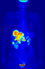
| ۱۱:۲۹, ۲۲ مې ۲۰۰۶ |  | ۲۰۰ × ۳۰۲ (۵۷۱ کيلوبايټ) | Damato | {{Information| |Description=Multi Intensity Projection PET image |Source=own work |Date=22. Mai 2006 |Author=Jens Langner |Permission=Public Domain }} |
د دوتنې کارېدنه
دا لاندينی مخ د همدې دوتنې سره تړنې لري:
د نړېوالې دوتنې کارېدنه
همدا دوتنه لاندينۍ نورې ويکي گانې کاروي:
- په ar.wikipedia.org کارونې
- په az.wikipedia.org کارونې
- په bg.wikipedia.org کارونې
- په ca.wikipedia.org کارونې
- په de.wikipedia.org کارونې
- په de.wikibooks.org کارونې
- په en.wikipedia.org کارونې
- Positron emission tomography
- Nuclear medicine
- Scientific visualization
- History of neuroimaging
- Talk:Nuclear medicine
- Portal:Medicine
- Fluorodeoxyglucose (18F)
- User talk:Damato
- User:Sbharris
- Wikipedia:Featured pictures/Sciences/Biology
- Radioactivity in the life sciences
- Spinning dancer
- Fluorine
- User:JerkerES
- User talk:Nergaal/Archive 5
- Wikipedia:WikiProject Medicine/Recognized content
- Wikipedia:Featured pictures thumbs/25
- Wikipedia:Featured picture candidates/October-2010
- Wikipedia:Featured picture candidates/Positron Emission Tomography
- Wikipedia:Featured picture candidates/new layout
- Wikipedia:Featured picture candidates/new layout b
- Wikipedia:Wikipedia Signpost/2010-10-25/Features and admins
- User:Public Juju/FP
- User:Laurenferruccio/sandbox
- Template:POTD/2012-07-04
- Temporal dynamics of music and language
- Talk:Science/Archive 6
- Biological aspects of fluorine
- User:Wouterstomp/test
- Ligand binding assay
- Wikipedia:Wikipedia Signpost/Single/2010-10-25
- Sandip Basu
د دې دوتنې نورې نړېوالې کارېدنې کتل.